Blog

Nov 12 2018

Reliable and Predictive In Vitro Assays for Myelotoxicity and Cardiotoxicity of Kinase Inhibitors

Clarke E1., Schwengberg S2., Kettenhofen R2., dos Santos G1., Bohlen H2., Society of Toxicology 48th Annual Meeting & ToxExpo 2009, Poster Abstract No.1854. 1 ReachBio Research Labs, Seattle, WA, USA,  2 Axiogenesis AG, Cologne, Germany

 INTRODUCTION

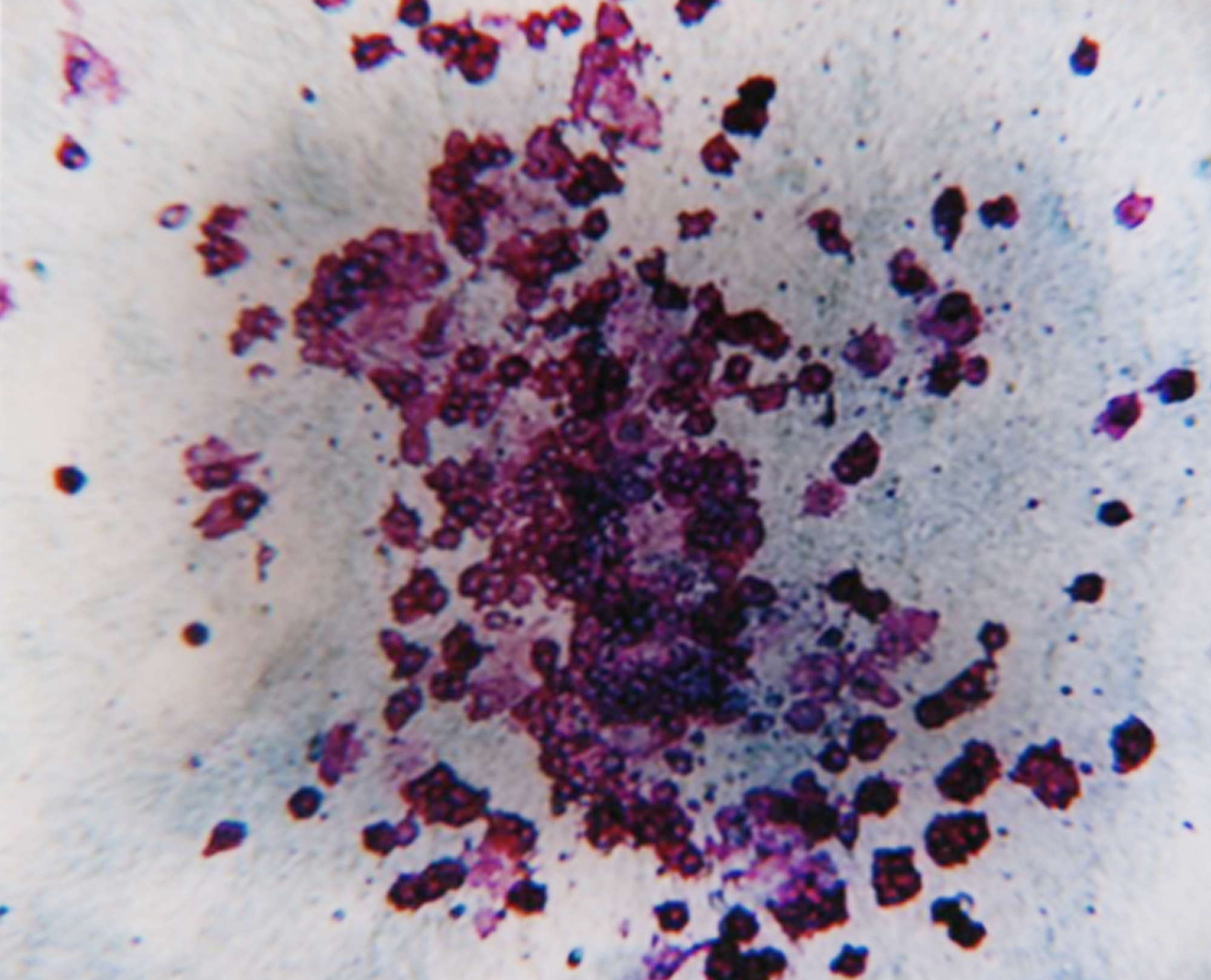
Kinase inhibitors (KIs) represent a new class of rationally designed drugs. The success of Imatinib, targeting the ABL tyrosine kinase in CML, has prompted the development of other KIs for the treatment of various cancers and inflammation. Although more successful than conventional therapies, myelotoxicity and cardiotoxicity are often major side effects of KIs. In order to predict if newly developed molecules demonstrated significant myelotoxicity or cardiotoxicity, we assessed a number of TKIs using in vitro models. To assess myelotoxicity, a human bone marrow progenitor assay (colony forming cell or CFC assay) previously validated by ECVAM was used. To assess cardiotoxicity, mouse ES cell-derived cardiomyocytes were used as a primary-like cellular platform in an in vitro cytotoxicity assay.


MATERIALS AND METHODS

For the in vitro myelotoxicity assay, clonogenic progenitors of the human myeloid (CFU-GM) lineage were set up in the methylcellulose-based media formulation (R&D Systems, MN). Six KIs were selected for testing: Imatinib, Lapatinib, Erlotinib, Dasatinib, Sorafenib and Sunitinib. These were chosen based upon their different target and disease specifications (Table 1) as well as reported differences in their clinical toxicity profiles. The KIs were added to the medium to give final concentrations ranging from 100 to 0.001 µg/mL. Solvent control cultures were also initiated. The cultures were set up in triplicate with normal pre-qualified human bone marrow (ReachBio, WA) from three different donors. Following incubation at 37°C, 5% CO2, for 14-16 days, the colonies were assessed and scored by trained personnel.

For the in vitro cardiotoxicity assay, mouse ES cell-derived cardiomyocytes (Cor.AT® cells, Axiogenesis, Germany) were plated in 24-well plates at 20,000 cells per well (6 replicates per condition) in Cor.At culture medium. The cells were cultured for 14 days so as to obtain a mature phenotype. The KIs were then added at concentrations ranging from 10-4 – 10-9 M. Cell death was measured by neutral red uptake 48 hours after the addition of KIs. The KIs were also added for the same amount of time and at the same concentrations used with the cardiomyocyte cultures to cultures of mouse embryonic fibroblasts (used as non-specific control cells), and fibroblast cell death was assessed by neutral red uptake.


Table 1 Target and Disease Specifications

TKI TARGET TREATMENT
Imantinbin ABL/PDGF/KIT CML and Ph+ B-ALL
Desatinib ABL/PDGF/KIT/src CML
Sorafenib VEGFR2/KIT/PDGF/RAF/FLt3 Renal cell carcinoma/myeloma
Sunitinb VEGFR1-3/KIT/PDGF/CSFR1/FLt3 Renal cell carcinoma
Lapatinib ERB/EGFR Breast Cancer
Erotinib RGFR/mutant JAK2 kinase Lung Cancer and possibly PV, MF


RESULTS

Rank order of myelotoxiciity potential in vitro correlates with published clinical observations.

The IC50 values derived from the in vitro CFC assays allowed us to rank the CFCs in terms of myelotoxicity (Figure 2). Remarkably, there was a direct correlation between the reported clinical myelotoxicity and the IC50 values derived from the CFC assays, with lower IC values associated with increased neutropenia (Table 2, Figure 1). Dasatinib was the most toxic KI (IC50 value: 0.008 ug/mL) and Lapatinib was the least toxic (IC50 value > 100 ug/mL), Table 2, Figure 2).

In vitro cardiotoxicity assay results and clinical observations closely associated.

There was also a strong association between published clinical cardiotoxicity (e.g. myopericarditis, ischemia, pericarditis, myocardial infarction) and the in vitro effect of the KIs on mature (day 14) cultures of cardiomyocytes derived from mouse ES cells (Cor.At® cells) (Table 3). Sorafenib was the most toxic KI where as Erlotinib and Lapatinib demonstrated the least toxicity (Table 3 and Figure 3). The profile of toxicity of the KIs on mouse ES-derived cardiomyocytes was not the same as that from mouse embryonic fibroblasts, suggesting that specific cardiomyocyte toxicity could be distinguished from a more generalized cytotoxic effects in these assays (Table 3).


Figure 1 Correlation between CFU-GM IC50 Values and Clinical Neutropenia for Kinase Inhibitors

 SOT09-f1


Figure 2  Kinase Inhibitor Myelotoxicity may be Ranked, based on CFU-GM Assay

SOT09-f2


Table 2 In Vitro CFU-GM IC50 Values Correlate with Clinical Neutropenia

  TOXICITY RANKING IC50 CFU-GM NEUTROPENIA GRADE 1/11 NEUTROPENIA GRADE 111/1V REFERENCE
Dasatinib 1 0.008 ug/mL 30% 33% J Clin Oncol 26 (19): 3204-3212, 2008
Sunitinib 2 0.09 ug/mL 32% 13% J Clin Oncol 24 (1): 16-24, 2006
Imatinib 3 2.6 ug/mL 6% 2% J Clin Oncol 22 (1): 77-85, 2004
Sorafenib 4 3.5 ug/mL ND 7% J Clin Oncol 25 (24): 3766-3773, 2006
Erlotinib 5 16 ug/mL ND ND J Clin Oncol 26 (4): 563-569, 2008
Lapatinib 6 >100 ug/mL ND ND The Oncologist 12: 756-765, 2007

  ND= none detected


Table 3 IC50 Values from an In Vitro Cardiomyocyte (Cor AT®) Toxicity Assay is Associated with Clinical Cardiotoxicity

  IC50 NEUTRAL RED UPTAKE ES_DERIVED CARDIOMYOCYTES IC50 NEUTRAL RED UPTAKE MOUSE EMBRYONIC FIBROBLASTS RANKING COR.AT/MEF CARDIOTOXICITY IN VITRO REFERENCE
 Sorafenib 0.229 μg/mL 12.6 μg/mL 1 ++ J Clin Oncol 24(9): 1363-1369, 2006
Imatinib 0.442 μg/mL 18.6 μg/mL 2 ++ Nat Med 12(8): 908-916, 2006
Dasatinib 0.203 μg/mL 4.54 μg/mL 3 ++  
Sunitinib 0.142 μg/mL 0.465 μg/mL 4 ++ Mol Pharmacol 74 (6): 1722-1728, 2008
Lapatinib 4.97 μg/mL 2.16 μg/mL 5 / The Oncologist 12:756-765, 2007
Erlotinib ND 0.528 μg/mL 6 / NA (only reports on skin and lung toxicity)

 ND= none detected

Figure 3 Comparison of Toxicity Profiles for Kinase Inhibitors in Cor At® Cardiomyocytes and Mouse Embryonic Fibroblasts

Figure3


 CONCLUSIONS

  • There is a direct correlation (r2 = 0.81) between the in vitro human CFU-GM IC50 values for various kinase inhibitors and clinical neutropenia.
  • The human CFU-GM assay can be used to rank KIs in terms of myelotoxicity potential as has been shown previously with certain other compound classes.
  • The human CFU-GM assay could be used to compare myelotoxicity between various classes of compounds and predict the myelotoxicity potential of new compounds.
  • There is an association between the results of the in vitro cytototoxicity assays using mouse ES-derived cardiomyocytes (Cor AT®) and clinical cardiotoxicity. Work is ongoing to optimize this assay on a number of different parameters and additional tests are being performed to study specific mechanisms of the
    toxicity in vitro (e.g., mitochondrial dysfunction).